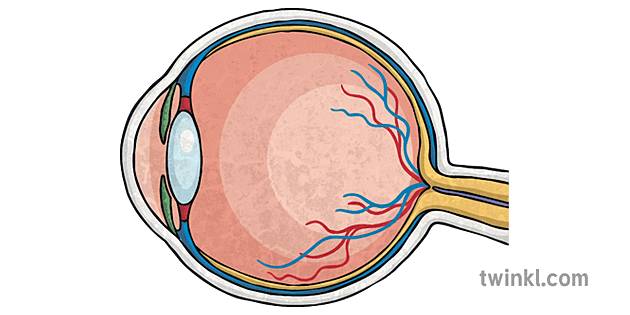
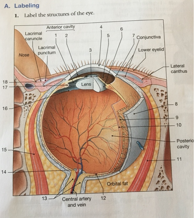
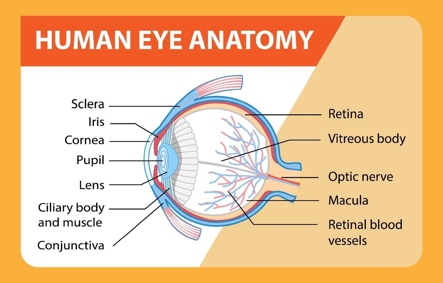
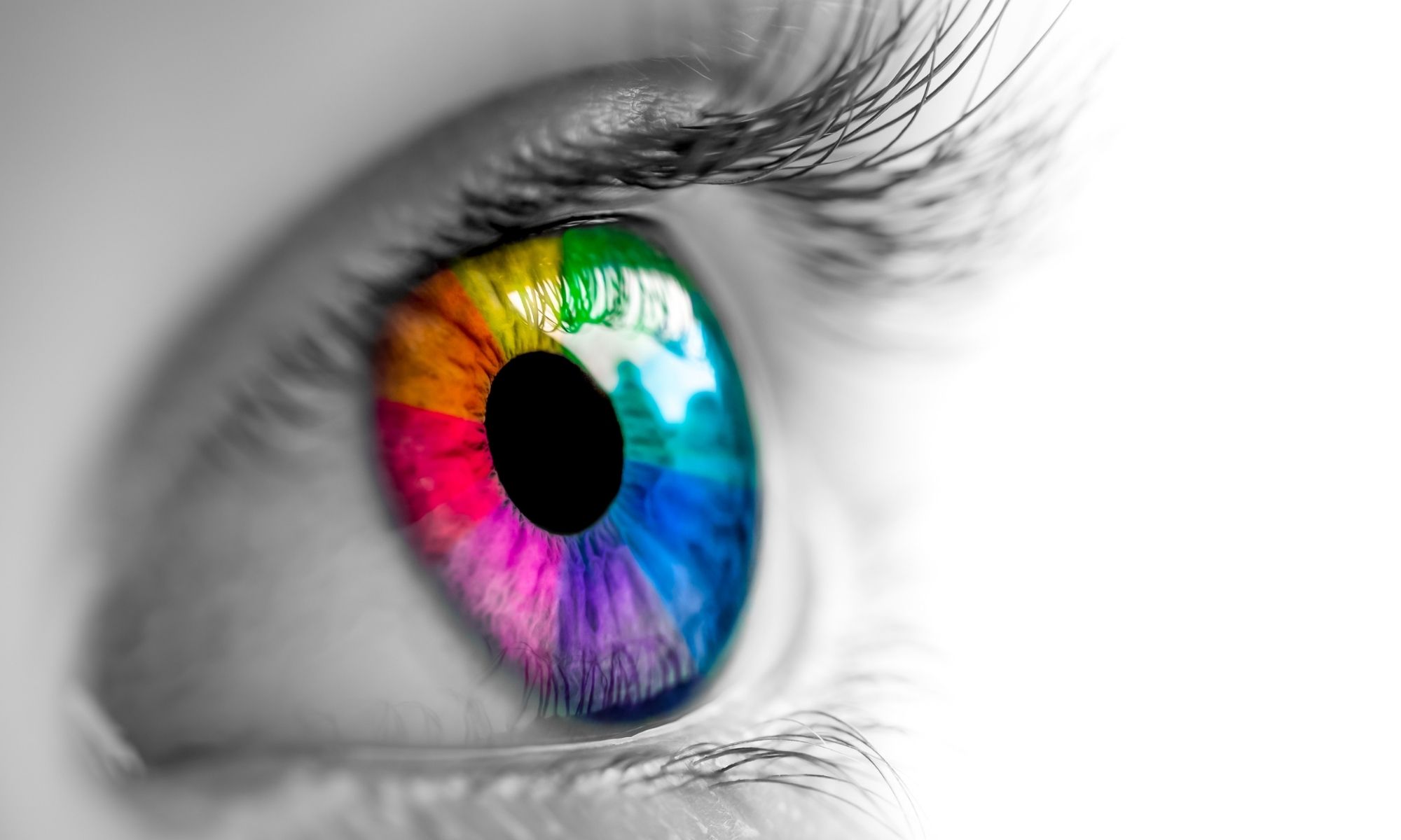
41 picture of the eye with labels
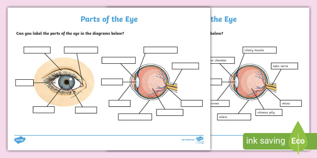
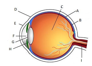
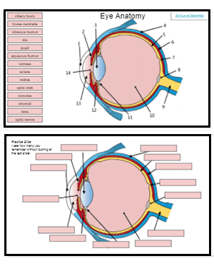
Solved B с A E F D Match the following parts of the eye with - Chegg Anatomy and Physiology questions and answers. B с A E F D Match the following parts of the eye with the labels in the picture above. A Iris F Cornea В. Ciliary Muscles G Optic Nerve C Lens E Retina Aqueous and Vitreous Fluid. Question: B с A E F D Match the following parts of the eye with the labels in the picture above. Label the Eye Worksheet - Teacher-Made Learning Resources - Twinkl In this resource, you'll find a 2-page PDF that is easy to download, print out, and use immediately with your class. The first page is a labelling exercise with two diagrams of the human eye. One is a view from the outside, and the other is a more detailed cross-section. Challenge learners to label the parts of the eye diagram. On the second page, you'll find a set of answers showing ...
Eye Pictures, Anatomy & Diagram | Body Maps - Healthline There are two types: cones make color vision possible, and rods specialize in black-and-white images. Although our eyes can only see in two dimensions, we are able to determine distances and depth...
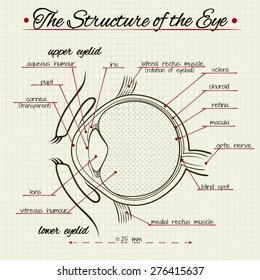
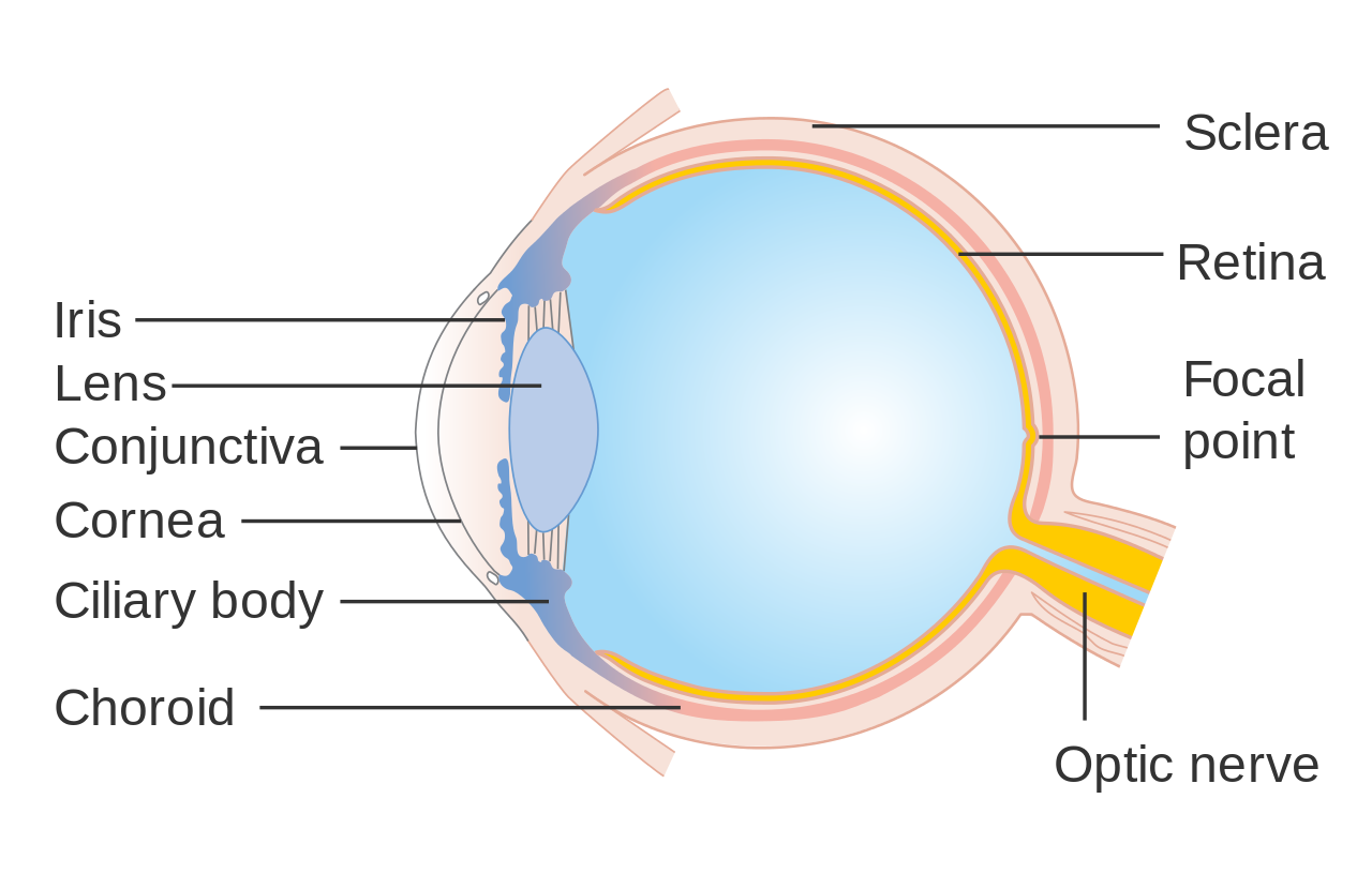
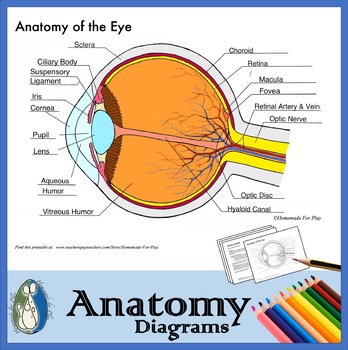
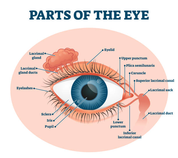
Picture of the eye with labels
PDF Eye Anatomy Handout - National Institutes of Health of light entering the eye. Lens: The lens is a clear part of the eye behind the iris that helps to focus light, or an image, on the retina. Macula: The macula is the small, sensitive area of the retina that gives central vision. It is located in the center of the retina. Optic nerve: The optic nerve is the largest sensory nerve of the eye. Dear Annie: Picture on phone has me worried about my husband ... Aug 23, 2022 · Dear Annie: Recently, I got married after being single and a widow for 23 years. My husband and I are in our late 70s and very active. We went on a tour to the Midwest. There was a very attractive ... Human Eye Anatomy Pictures, Images and Stock Photos Vector illustration of the structure of the eye. Anatomy of the... Structure of the eye, parts of the eye. Retina, macula, blind spot, optic nerve, cones, rods, vitreous humor, ciliary body, lens, pupil, aqueous humor, cornea, iris, sclera, choroid. Eye layers The inner layer of the eye, or retina, is similar to film in a camera.
Picture of the eye with labels. Eye Anatomy: 16 Parts of the Eye & Their Functions - Vision Center The lens of the eye (or crystalline lens) is the transparent lentil-shaped structure inside your eye. This is the natural lens. It is located behind the iris and to the front of the vitreous humor (vitreous body). The vitreous humor is a clear, colorless, gelatinous mass that fills the gap between the lens and the retina in the eye. Mpofu labels Samuel as an 'unrepentant liar' in Mkhwebane ... Aug 02, 2022 · Mpofu labels Samuel as an 'unrepentant liar' in Mkhwebane impeachment inquiry Busisiwe Mkhwebane’s lawyer, Dali Mpofu, said that Sphelo Samuel’s testimony before this inquiry has been born out ... Optic Nerve Photos and Premium High Res Pictures - Getty Images Browse 599 optic nerve stock photos and images available, or search for eye optic nerve or optic nerve damage to find more great stock photos and pictures. Related searches: eye optic nerve. optic nerve damage. optic nerve icon. Eye Anatomy Diagram - EnchantedLearning.com Definitions : Aqueous humor - the clear, watery fluid inside the eye. It provides nutrients to the eye. Astigmatism - a condition in which the lens is warped, causing images not to focus properly on the retina. Binocular vision - the coordinated use of two eyes which gives the ability to see the world in three dimensions - 3D.
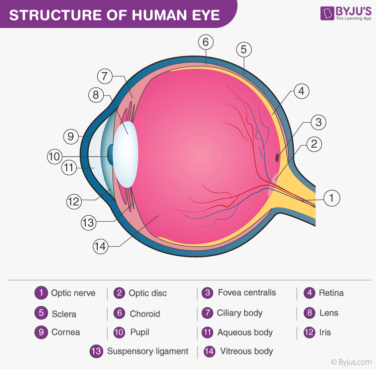
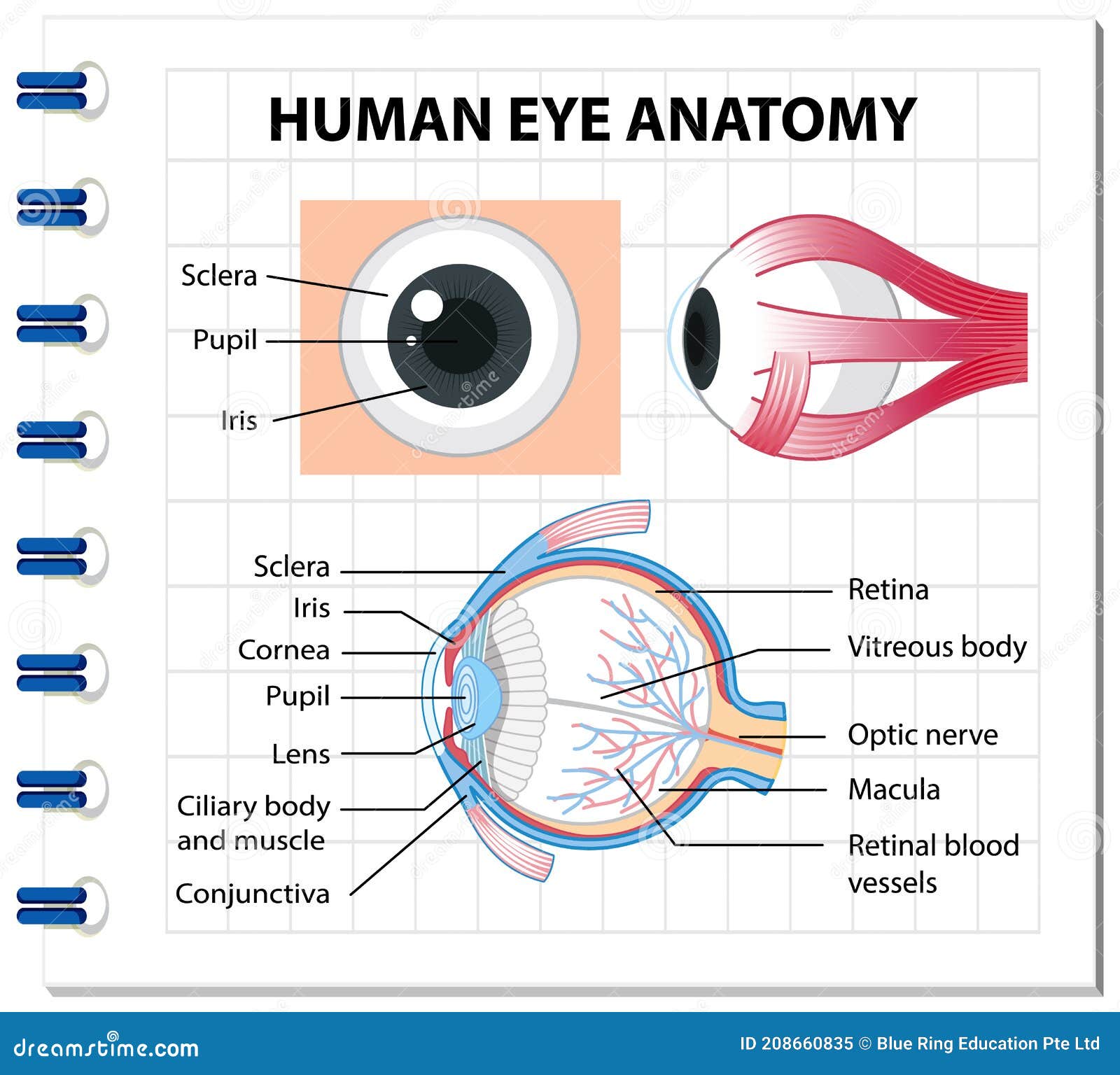
Motion Picture Association - Wikipedia The Motion Picture Association represents the interests of the six international producers and distributors of filmed entertainment. To do so, they promote and protect the intellectual property rights of these companies and conduct public awareness programs to highlight to movie fans around the world the importance of content protection. PDF Parts of the Eye - National Institutes of Health Eye Diagram Handout Author: National Eye Health Education Program of the National Eye Institute, National Institutes of Health Subject: Handout illustrating parts of the eye Keywords: parts of the eye, eye diagram, vitreous gel, iris, cornea, pupil, lens, optic nerve, macula, retina Created Date: 12/16/2011 12:39:09 PM Eye Diagram With Labels and detailed description - BYJUS A brief description of the eye along with a well-labelled diagram is given below for reference. Well-Labelled Diagram of Eye The anterior chamber of the eye is the space between the cornea and the iris and is filled with a lubricating fluid, aqueous humour. The vascular layer of the eye, known as the choroid contains the connective tissue. Eye Anatomy Detail Picture Image on MedicineNet.com Picture of Eye Anatomy Detail The eye is our organ of sight. The eye has a number of components which include but are not limited to the cornea, iris, pupil, lens, retina, macula, optic nerve, choroid and vitreous. Cornea: clear front window of the eye that transmits and focuses light into the eye.
Human Eye Diagram - Human Body Pictures & Images - Science for Kids Photo description: This human eye diagram gives an excellent overview of the human eye. The cross section features labeled parts such as the iris, pupil, cornea, lens, retina, choroid, optic disc, optic nerve and fovea. For more information on eyes, check out our range of interesting human eye facts. Diagram of the Eye - Lions Eye Institute Instructions Click the parts of the eye to see a description for each. Hover the diagram to zoom. Need any help? If you would like to know more about us, or want to make an appointment, please don't hesitate to get in touch. (08) 9381 0777 carecentre@lei.org.au Request an appointment Customer Care Centre (08) 9381 0777 Eye Anatomy: Parts of the Eye and How We See Behind the anterior chamber is the eye's iris (the colored part of the eye) and the dark hole in the middle called the pupil. Muscles in the iris dilate (widen) or constrict (narrow) the pupil to control the amount of light reaching the back of the eye. Directly behind the pupil sits the lens. The lens focuses light toward the back of the eye. Bunnie Xo - Biography - IMDb With all of her current success she doesn't plan on stopping anytime soon. She hopes to one day have her own radio show. As her current shows continue to grow at a exponential rate she keeps her eye on the prize and stays all gas no breaks. - IMDb Mini Biography By: Vivace Hub
12 Best Sticker Printer For Labels, Stickers, And Photos In 2022 Aug 07, 2022 · 10 sheets of 2×3 inch paper- for fast pictures or stickers, you may use the adhesive back picture paper with the peel-and-stick backing that comes with the kit. Features: Customize photos with borders and stickers, print from social media or your camera roll, view your photos in augmented reality, easily view photo libraries from the app ...
Quiz: Label The Parts Of The Eye - ProProfs Quiz Quiz: Label The Parts Of The Eye. Do you know the anatomy of the human eye very well? Can you label the parts of the eye in the quiz below? Give it a try and evaluate yourself. The eye has many important parts, each with different functions, including the cornea, pupil, sclera, and many more. Can you tell where these parts are located and what ...
Label Eye Printout - EnchantedLearning.com Label the Eye Diagram. Human Anatomy. Read the definitions, then label the eye anatomy diagram below. Cornea - the clear, dome-shaped tissue covering the front of the eye. Iris - the colored part of the eye - it controls the amount of light that enters the eye by changing the size of the pupil. Lens - a crystalline structure located just behind ...
Amazon.com: 50 Chalkboard Labels - Includes Erasable Chalk ... Mr. Pen- Chalkboard Labels, 100pc, Assorted Shapes, 1 White Chalk Marker and Small Towel, Labels, Label Stickers, Labels for Storage Bins, Sticker Labels, Bottle Labels, Food Labels, Jar Labels 4.7 out of 5 stars 1,679
31 Most Beautiful Eyes in the World - Woman's World We bet anyone that meets them feels the same way! Getty Images Eyes are naturally beautiful — from the delicate shapes and unique colors to the countless expressions that can be made with them. Blue eyes, brown eyes, green eyes, hazel eyes, gray eyes, and any shade in between are all stunning. Please don't ask us to pick a favorite!
Label Parts of the Human Eye - University of Dayton Parts of the Eye. Select the correct label for each part of the eye. The image is taken from above the left eye. Click on the Score button to see how you did. Incorrect answers will be marked in red. ...
Bunnie Xo - Biography - IMDb With all of her current success she doesn't plan on stopping anytime soon. She hopes to one day have her own radio show. As her current shows continue to grow at a exponential rate she keeps her eye on the prize and stays all gas no breaks. - IMDb Mini Biography By: Vivace Hub
Label Functions of Parts of the Human Eye - University of Dayton Select the correct label for the function of each part of the eye. The image is taken from above the left eye. Click on the Score button to see how you did. Incorrect answers will be marked in red.
Amazon.com: 2-Pack of 1000-Piece Jigsaw Puzzles, for Adults, … 15-07-2020 · Amazon.com: 2-Pack of 1000-Piece Jigsaw Puzzles, for Adults, Families, and Kids Ages 8 and up, Retro Comics and Fruit Labels, Amazon Exclusive : Everything Else
Return Address Labels & Envelope Seals - Miles Kimball These peel-and-stick return address labels are available in white, gold or silver to let the crisp black font stand out while adding elegance to your envelopes. Go the extra mile with these eye-catching labels just right for sending Christmas cards, personal letters, invites, office communication and so much more.
(Don't) look here!: The effect of different forms of label added to ... Participants were 260 female undergraduate students whose eye movements were recorded while viewing three thin-ideal fashion advertisements with one of five different forms of label added: no label, disclaimer label (indicating image had been digitally altered), consequence label (indicating that viewing images might make women feel bad about ...
What is an eye mark and why do I need it? - Consolidated Label An 'eye mark' (also known as 'eye spot') is a small rectangular printed area located near the edge of the printed flexible packaging material. A sensor on the form-fill-seal (FFS) machine reads the eye mark to identify packaging material, control the material's position, and coordinate the separation and cutting of the flexible packaging material.
1,111,045 Human eye Images, Stock Photos & Vectors - Shutterstock 1,111,045 human eye stock photos, vectors, and illustrations are available royalty-free. See human eye stock video clips Image type Orientation Artists Sort by Popular Biology Healthcare and Medical Icons and Graphics human eye macro photography anatomy iris eye 3d rendering pupil Next of 11,111
Labelling the eye — Science Learning Hub In this interactive, you can label parts of the human eye. Use your mouse or finger to hover over a box to highlight the part to be named. Drag and drop the text labels onto the boxes next to the eye diagram If you want to redo an answer, click on the box and the answer will go back to the top so you can move it to another box.
The Human Eye (Eyeball) Diagram, Parts and Pictures The eyeball is a round gelatinous organ that contains the actual optical apparatus. It is approximately 25 mm in diameter and sits snugly in the orbit where six muscles control its movement. The eyeball has three layers, each of which has several important structures that are essential for the sense of vision. Wall of the Eyeball
30 Eye-Catching Wine Label Designs For Inspiration The designer used squares to provide the information. 02. The Cloud Factory. The Cloud Factory wine label design looks simple because of its use of two colors only. Yellow and white colors give the label a soft look, which seems to be the intention of the designer and the brand. 03.
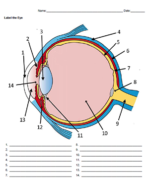
Label the Eye - The Biology Corner Label the Eye Shannan Muskopf December 30, 2019 This worksheet shows an image of the eye with structures numbered. Students practice labeling the eye or teachers can print this to use as an assessment. There are two versions on the google doc and pdf file, one where the word bank is included and another with no word bank for differentiation.
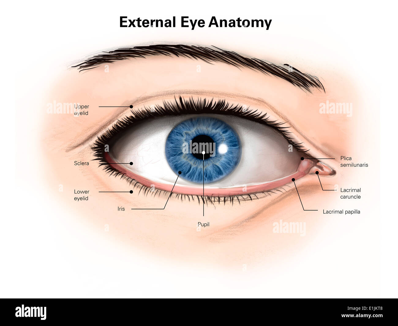
WebMD - Better information. Better health. Your eye is a slightly asymmetrical globe, about an inch in diameter. The front part (what you see in the mirror) includes: Iris: the colored part. Cornea: a clear dome over the iris. Pupil: the ...
Transverse section of eye anatomy with labels. - Getty Images Transverse section of eye anatomy with labels. - stock illustration. Transverse section of eye anatomy with labels. Buy the print. PURCHASE A LICENSE. All Royalty-Free licenses include global use rights, comprehensive protection, simple pricing with volume discounts available.
540,440 Eye drawing Images, Stock Photos & Vectors - Shutterstock Eye drawing royalty-free images. 540,440 eye drawing stock photos, vectors, and illustrations are available royalty-free. See eye drawing stock video clips. Image type.
Human Eye Anatomy Pictures, Images and Stock Photos Vector illustration of the structure of the eye. Anatomy of the... Structure of the eye, parts of the eye. Retina, macula, blind spot, optic nerve, cones, rods, vitreous humor, ciliary body, lens, pupil, aqueous humor, cornea, iris, sclera, choroid. Eye layers The inner layer of the eye, or retina, is similar to film in a camera.
Dear Annie: Picture on phone has me worried about my husband ... Aug 23, 2022 · Dear Annie: Recently, I got married after being single and a widow for 23 years. My husband and I are in our late 70s and very active. We went on a tour to the Midwest. There was a very attractive ...
PDF Eye Anatomy Handout - National Institutes of Health of light entering the eye. Lens: The lens is a clear part of the eye behind the iris that helps to focus light, or an image, on the retina. Macula: The macula is the small, sensitive area of the retina that gives central vision. It is located in the center of the retina. Optic nerve: The optic nerve is the largest sensory nerve of the eye.
:background_color(FFFFFF):format(jpeg)/images/library/11201/overview_parts_of_the_eye_labelled_diagram.jpg)









(249).jpg)






Post a Comment for "41 picture of the eye with labels"