38 human skull diagram with labels
Blood vessels of the head and neck - Anatomy and Physiology The majority of blood draining from the head is passed into three pairs of veins: • vertebral veins. Within the brain all veins lead to the internal jugular veins. The external jugular veins are smaller than the internal jugular veins and lie superficial to them. They receive blood from superficial regions of the face, scalp and neck. Labeling Leg Muscles - Human Anatomy - GUWS Medical 1. Review textbook sections on muscles that move the thigh, muscles that move the leg, and muscles that move the foot. 2. As a review activity, label figures 23.1, 23.2, 23 3, 23.4, 23.5, and 23.6. 3. Locate the following muscles in the torso and in the lower limb models. Also locate as many of them as possible in your own body.
Organs of the body | Their Locations and Internal Functions This is the master organ of the body. All the organ systems of the human body are under its control. The skull, a bone frame in the head, houses the brain. It is made up of nerve cells and neuroglia. It consists of parts like the cortex, cerebral hemisphere, cerebellum, medulla oblongata, and pons. It extends into the spinal cord.
Human skull diagram with labels
Appendicular Skeleton: Bones List, Diagram & More - Embibe The pelvic girdle is a composition of bones that functions as the point of attachment of the lower limbs to the axial skeleton. It comprises two coxal bones or hip bones. Coxal bone (hip bones) is also called ossa coxae or innominate bone . Each coxal bone comprises three fused bones: the upper ilium, the lower ischium, and the inner pubis. Organs of Skeletal System and Their Functions - New Health Advisor 1. Bones. The most important organ of the skeletal system is the bones. Human skeleton is made up of 206 bones that in coordination not only provides support and protection to the viscera (with the help of muscles attached to them) but also produces blood cells for the body from the bone-marrow. 2. Posterior view of skeleton illustrations and clipart (183) Set of Anatomically correct realistic flat Clip Art by Katya_Golovchyn 0 / 0 Skeleton Human diagram front back anterior posterior view with two arm poses. ... Stock Illustration by GoodStudio 0 / 0 Skeleton Pelvis Posterior view Stock Illustrations by sciencepics 0 / 2,258 Human Skeleton System Rib Cage with Labels Anatomy ...
Human skull diagram with labels. Gross Anatomy Of The Skeletal Muscles Answers considered, human muscle—like the muscles of all vertebrates—is often divided into striated muscle (or skeletal muscle), smooth muscle, and cardiac muscle.Smooth muscle is under involuntary control... Human brain - Wikipedia The cerebrum, the largest part of the human brain, consists of two cerebral hemispheres. Each hemisphere has an inner core composed of white matter, and an outer surface - the cerebral cortex - composed of grey matter. The cortex has an outer layer, the neocortex, and an inner allocortex. Skull: Foramina, fissures and contents - Kenhub Foramen caecum (or cecum): Is the most anterior of the holes in the floor of the skull. It lies in the frontal bone, just anterior to the ethmoid bone. It allows the passage of an emissary vein that comes from the nasal cavity and drains into the superior sagittal sinus, part of the venous drainage system associated with the brain. Anatomical Line Drawings - Medscape Surface Anatomy - lateral views - male. go to drawing without labels. Surface Anatomy - lateral views - female. go to drawing without labels. Surface Anatomy - Child - anterior view & posterior ...
Diagram of Human Heart and Blood Circulation in It A heart diagram labeled will provide plenty of information about the structure of your heart, including the wall of your heart. The wall of the heart has three different layers, such as the Myocardium, the Epicardium, and the Endocardium. Here's more about these three layers. Epicardium Diagram Longbone Image - Graph Diagram This human anatomy diagram with labels depicts and explains the details and or parts of the Diagram Longbone Image. Human anatomy diagrams and charts show internal organs, body systems, cells, conditions, sickness and symptoms information and/or tips to ensure one lives in good health. ... such as the epiphysis. Blank Skull Diagram - Blank ... Axial Skeleton Anatomy: Diagram, Definition, Functions - Embibe The axial skeleton is the section of a vertebrate's skeleton that comprises the head and trunk bones. The human skeleton is made up of 80 bones and is divided into six sections: the skull (22 bones), middle ear ossicles, hyoid bone, rib cage, sternum, and spinal column. The axial and appendicular skeletons combine to produce the entire skeleton. Sutures of the skull: Anatomy | Kenhub Check out our skull bones quizzes and diagrams. Posterior view The sagittal suture joins the two parietals. The lambdoid suture marks the borders between the parietal and occipital bones. The sagittal and lambdoid sutures converge into a lambda. Superior view The coronal suture separates the frontal bone and the parietal bone.
Anatomy Project - Sheridan College Neck. · Connecting the shaft and head of the femur. · Projects superior and medial from the shaft to the head. · In addition to projecting superior and medial from the shaft of the femur, the neck also projects somewhat anterior. · The amount of forward projection is extremely variable, but on an average is from 12° to 14°. WHMIS 2015 - Pictograms : OSH Answers The skull and crossbones pictogram is used for the following classes and categories: Acute toxicity - ... The product label and Section 2 (Hazards Identification) of the SDS still require the signal word, hazard statement(s), and other required label elements. WHMIS 2015 classes and categories that do not require a pictogram are: Incredible Skeleton Coloring Pages Anatomy 2022 - Ellison Blog The appendicu0cr skeleton, the concludes c discussion of the five types of bones making up the skeleton. Skeleton Coloring Printable Anatomy Cool2Bkids Skull Human Skeletons Bones Sheets Adult. An illustration of a skeleton with flowery eyes holding a pumpkin in the graveyard with bats flying in the night sky. Skull: Anatomy, structure, bones, quizzes | Kenhub The human skull consists of 22 bones (or 29, including the inner ear bones and hyoid bone) which are mostly connected together by ossified joints, so called sutures. The skull is divided into the braincase ( neurocr anium) and the facial skeleton ( viscerocranium ).
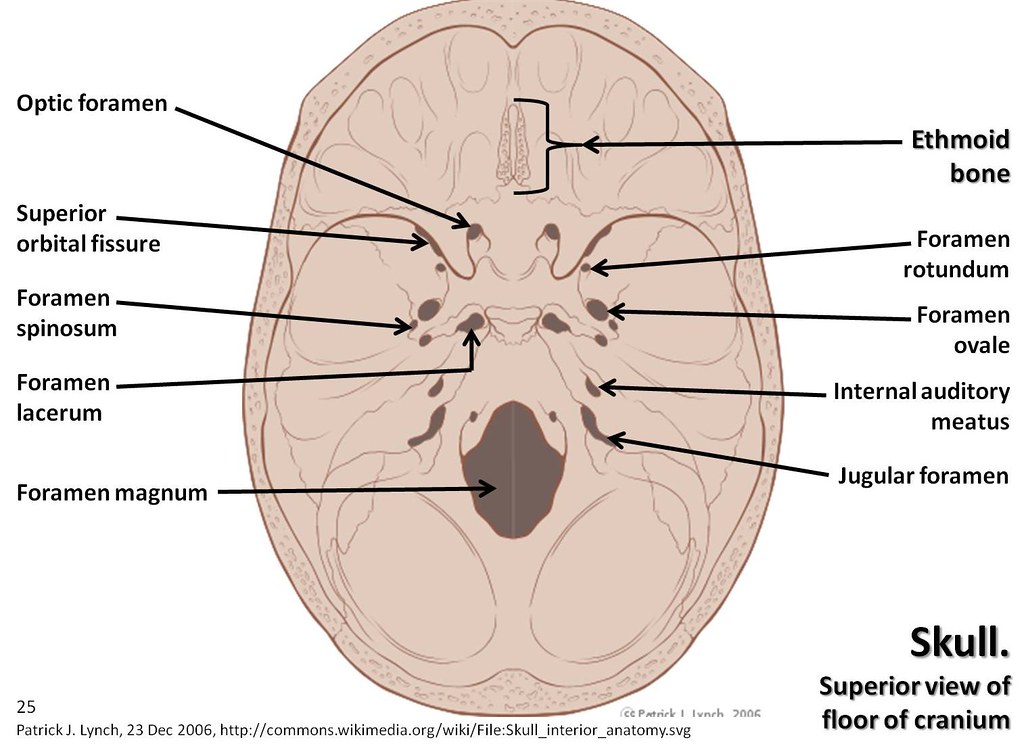
Skull diagram, superior view of floor of cranium with labe… | Flickr
How to Work Safely with - Hazardous Products using the "Skull and ... The symbol within the pictogram is a human skull with two crossed bones behind it. The symbol indicates that hazardous products with this pictogram can cause death or poisoning. Hazardous products with this pictogram can be safely worked with if proper storage and handling practices are followed.

Vector Illustration Of Diagram Of Human Skull - 187162106 : Shutterstock
Positions and Functions of the Four Brain Lobes - MD-Health.com The brain is divided into four sections, known as lobes (as shown in the image). The frontal lobe, occipital lobe, parietal lobe, and temporal lobe have different locations and functions that support the responses and actions of the human body. Let's start by identifying where each lobe is positioned in the brain. Position of the Lobes

Numbered Classic Human Skull - Anatomy Models and Anatomical Charts
Anatomy of the Human Shoulder Joint - Verywell Health The three bones of the shoulder are the: Humerus (arm bone) Scapula (shoulder blade) Clavicle (collarbone) The scapula has one part that forms a socket for the ball-and-socket shoulder joint; this is called the glenoid. The glenoid is covered with smooth cartilage.

Human Skeleton Blank Clip Art at Clker.com - vector clip art online, royalty free & public domain
Inferior view of the base of the skull: Anatomy | Kenhub In the middle section of the skull is the foramen Lacerum, which is a jagged opening that is filled with cartilage in life. It lies directly above the ICA ( internal carotid artery) during its course through the carotid canal. It can be described as the window through which the ICA can be seen (from above).

My Class Blog: Human Biology: Unit 3 Compilation
What are the 12 cranial nerves? Functions and diagram Scientists use Roman numerals from I to XII to label the cranial nerves in the brain. The 12 cranial nerves include the: olfactory nerve optic nerve oculomotor nerve trochlear nerve trigeminal...

Human Body Anatomy Basics No Lines Clip Art at Clker.com - vector clip art online, royalty free ...
Skull anatomy: Anterior and lateral views of the skull - Kenhub The human skull consists of about 22 to 30 single bones which are mostly connected together by ossified joints, so called sutures. The skull is divided into the braincase ( cerebral cranium) and the face ( visceral cranium ). The main task of the skull is the protection of the most important organ in the human body: the brain.

345862 Human Skull Diagram Anatomy Educational Chart Mural GLOSSY POSTER US | eBay
List of skeletal muscles of the human body - Wikipedia This is a table of skeletal muscles of the human anatomy.. There are around 650 skeletal muscles within the typical human body. Almost every muscle constitutes one part of a pair of identical bilateral muscles, found on both sides, resulting in approximately 320 pairs of muscles, as presented in this article. Nevertheless, the exact number is difficult to define.

'Anterior View of Human Skull, with Labels' Print - Stocktrek Images | AllPosters.com
CoordinateSystems - Brainstorm The nasion is the intersection of the frontal and two nasal bones of the human skull. Its manifestation on the visible surface of the face is a distinctly depressed area directly between the eyes, ... Volume of integers where each value represents an anatomical label, and registered to an MNI space. A non-linear deformation field can be applied ...
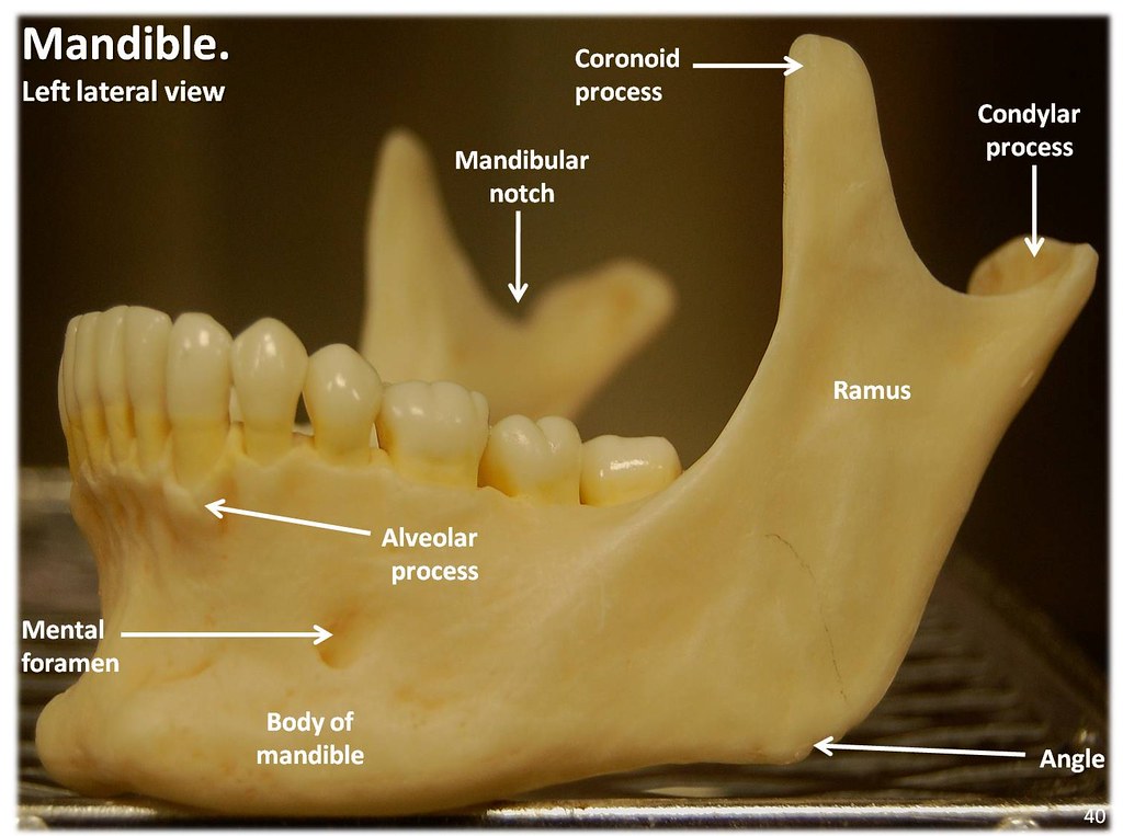
Mandible, lateral view with labels - Axial Skeleton Visual… | Flickr
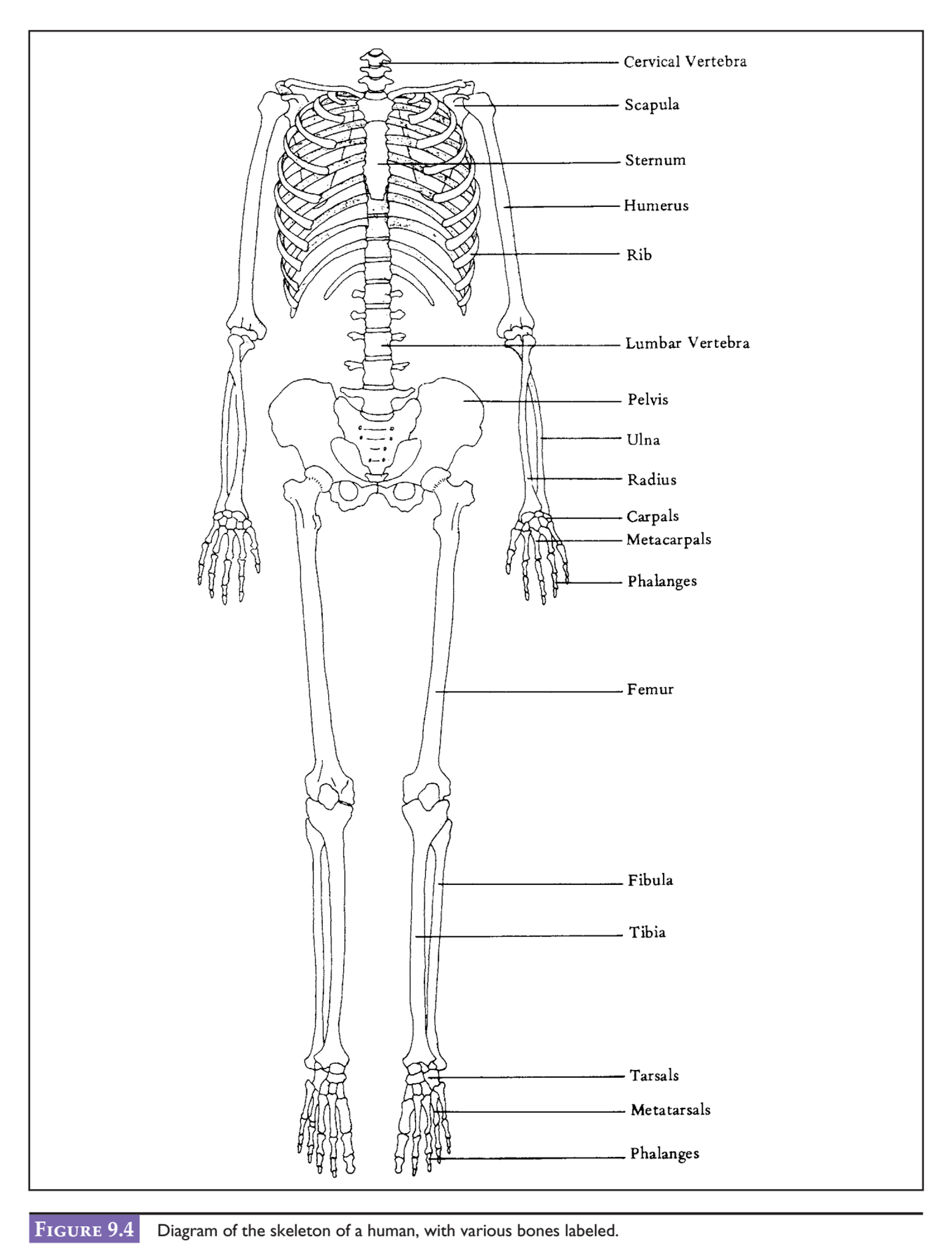
What's Inside Your Bones? - Lesson - TeachEngineering The human body is composed of 206 bones. Most are located in the hands (54) and feet (52). In the head alone are 29 bones: eight cranial bones, 14 facial bones, six ear bones and one throat bone. Below the head are four shoulder bones, 25 thorax bones and 24 vertebral column bones. In the arms are two upper arm bones and four forearm bones.
Skeleton Posterior Clip Art at Clker.com - vector clip art online, royalty free & public domain
Strange Science: Hominids This diagram of species diversity appeared in an early-20th-century biology textbook. If a similar diagram were published today, the species numbers would certainly be different. Some of the categories and relationships between organisms would be different, too. But the diagram gave a reasonable assessment of biological discoveries up to that time.

Diagram Of Human Skull Digital Art by Graphicaartis
Posterior view of skeleton illustrations and clipart (183) Set of Anatomically correct realistic flat Clip Art by Katya_Golovchyn 0 / 0 Skeleton Human diagram front back anterior posterior view with two arm poses. ... Stock Illustration by GoodStudio 0 / 0 Skeleton Pelvis Posterior view Stock Illustrations by sciencepics 0 / 2,258 Human Skeleton System Rib Cage with Labels Anatomy ...
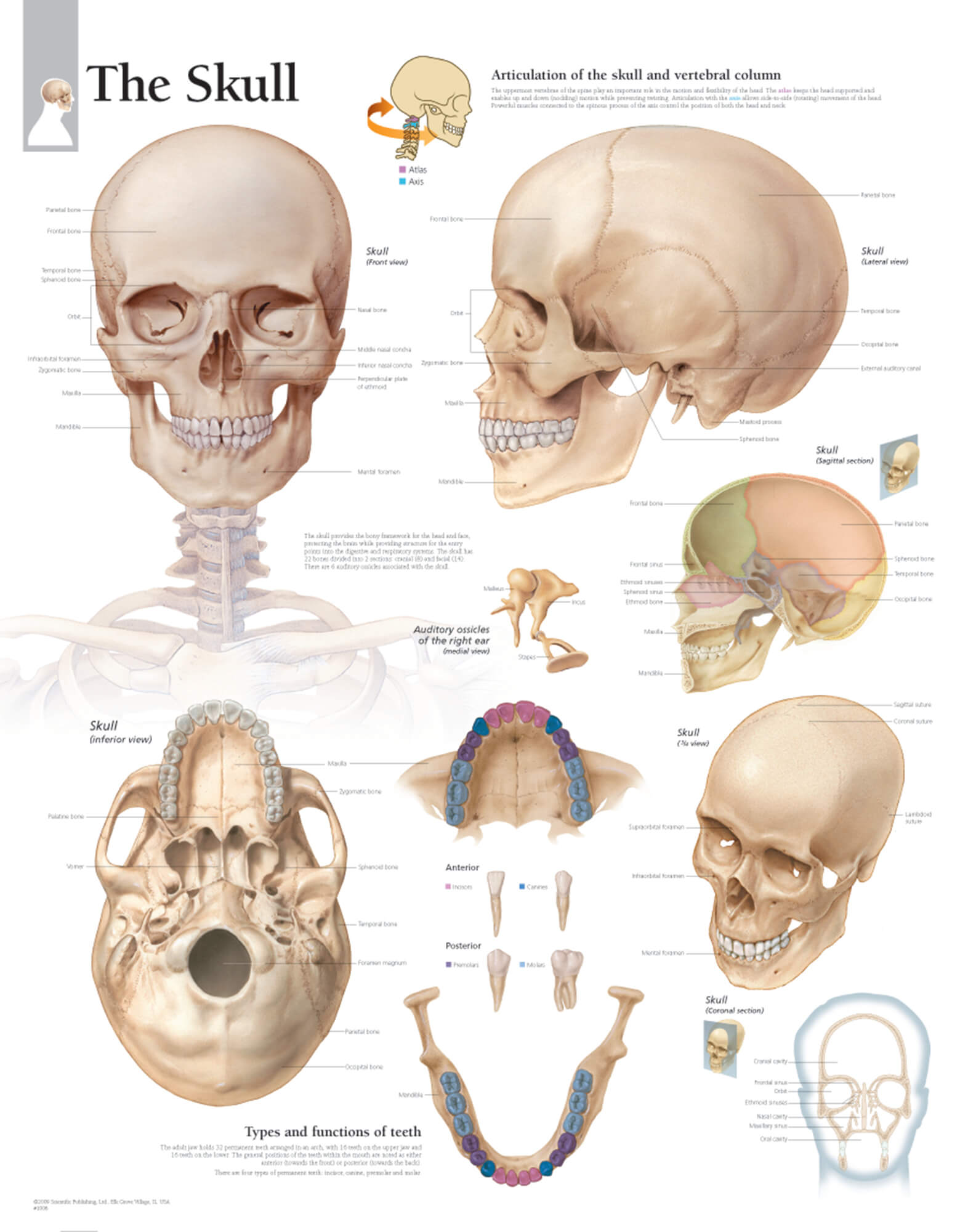
The Skull – Scientific Publishing
Organs of Skeletal System and Their Functions - New Health Advisor 1. Bones. The most important organ of the skeletal system is the bones. Human skeleton is made up of 206 bones that in coordination not only provides support and protection to the viscera (with the help of muscles attached to them) but also produces blood cells for the body from the bone-marrow. 2.

Blank Skull Bones Quiz Clip Art at Clker.com - vector clip art online, royalty free & public domain
Appendicular Skeleton: Bones List, Diagram & More - Embibe The pelvic girdle is a composition of bones that functions as the point of attachment of the lower limbs to the axial skeleton. It comprises two coxal bones or hip bones. Coxal bone (hip bones) is also called ossa coxae or innominate bone . Each coxal bone comprises three fused bones: the upper ilium, the lower ischium, and the inner pubis.
Solved: After Examining The Diagram Of A Human Skeleton In... | Chegg.com

human skeleton labeled

NEW THE HUMAN SKULL ANATOMY ANATOMICAL DIAGRAM GUIDE CHART PRINT PREMIUM POSTER | eBay
Post a Comment for "38 human skull diagram with labels"